Agarose Gel Electrophoresis
From 2007.igem.org
| (4 intermediate revisions not shown) | |||
| Line 1: | Line 1: | ||
It is a method used in biochemistry and molecular biology to separate DNA, RNA, or protein molecules by size. This is achieved by moving negatively charged nucleic acid molecules through an agarose matrix with an electric field (electrophoresis). | It is a method used in biochemistry and molecular biology to separate DNA, RNA, or protein molecules by size. This is achieved by moving negatively charged nucleic acid molecules through an agarose matrix with an electric field (electrophoresis). | ||
| - | + | Fragments of linear DNA migrate through agarose gels with a mobility that is inversely proportional to the log10 of their molecular weight. In other words, if you plot the distance from the well that DNA fragments have migrated against the log10 of either their molecular weights or number of base pairs, a roughly straight line will appear. | |
| - | + | Circular forms of DNA migrate in agarose distinctly differently from linear DNAs of the same mass. Typically, uncut plasmids will appear to migrate more rapidly than the same plasmid when linearized. Additionally, most preparations of uncut plasmid contain at least two topologically-different forms of DNA, corresponding to supercoiled forms and nicked circles. The image to the right shows an ethidium-stained gel with uncut plasmid in the left lane and the same plasmid linearized at a single site in the right lane. | |
| - | + | ||
| - | + | ||
| - | + | Several additional factors have important effects on the mobility of DNA fragments in agarose gels, and can be used to your advantage in optimizing separation of DNA fragments. Chief among these factors are: | |
| - | + | Agarose Concentration: By using gels with different concentrations of agarose, one can resolve different sizes of DNA fragments. Higher concentrations of agarose facilite separation of small DNAs, while low agarose concentrations allow resolution of larger DNAs. | |
| - | + | ||
| - | + | The image to the right shows migration of a set of DNA fragments in three concentrations of agarose, all of which were in the same gel tray and electrophoresed at the same voltage and for identical times. Notice how the larger fragments are much better resolved in the 0.7% gel, while the small fragments separated best in 1.5% agarose. The 1000 bp fragment is indicated in each lane. | |
| + | |||
| - | + | Voltage: As the voltage applied to a gel is increased, larger fragments migrate proportionally faster that small fragments. For that reason, the best resolution of fragments larger than about 2 kb is attained by applying no more than 5 volts per cm to the gel (the cm value is the distance between the two electrodes, not the length of the gel). | |
| - | + | ||
| - | + | ||
| - | + | Electrophoresis Buffer: Several different buffers have been recommended for electrophoresis of DNA. The most commonly used for duplex DNA are TAE (Tris-acetate-EDTA) and TBE (Tris-borate-EDTA). DNA fragments will migrate at somewhat different rates in these two buffers due to differences in ionic strength. Buffers not only establish a pH, but provide ions to support conductivity. If you mistakenly use water instead of buffer, there will be essentially no migration of DNA in the gel! Conversely, if you use concentrated buffer (e.g. a 10X stock solution), enough heat may be generated in the gel to melt it. | |
| - | + | Effects of Ethidium Bromide: Ethidium bromide is a fluorescent dye that intercalates between bases of nucleic acids and allows very convenient detection of DNA fragments in gels, as shown by all the images on this page. As described above, it can be incorporated into agarose gels, or added to samples of DNA before loading to enable visualization of the fragments within the gel. As might be expected, binding of ethidium bromide to DNA alters its mass and rigidity, and therefore its mobility. | |
| - | + | ||
| - | + | ||
| - | + | ||
| - | + | ||
| - | [ | + | [[Image:Gel.jpg|250px]] [[Image:Gel2.jpg|250px]] |
| - | + | ||
| - | + | ||
| - | + | ||
| - | + | ||
| - | + | ||
| - | + | ||
| - | + | ||
| - | + | ||
| - | + | ||
| - | + | ||
| - | + | ||
| - | + | ||
Latest revision as of 11:06, 21 October 2007
It is a method used in biochemistry and molecular biology to separate DNA, RNA, or protein molecules by size. This is achieved by moving negatively charged nucleic acid molecules through an agarose matrix with an electric field (electrophoresis). Fragments of linear DNA migrate through agarose gels with a mobility that is inversely proportional to the log10 of their molecular weight. In other words, if you plot the distance from the well that DNA fragments have migrated against the log10 of either their molecular weights or number of base pairs, a roughly straight line will appear.
Circular forms of DNA migrate in agarose distinctly differently from linear DNAs of the same mass. Typically, uncut plasmids will appear to migrate more rapidly than the same plasmid when linearized. Additionally, most preparations of uncut plasmid contain at least two topologically-different forms of DNA, corresponding to supercoiled forms and nicked circles. The image to the right shows an ethidium-stained gel with uncut plasmid in the left lane and the same plasmid linearized at a single site in the right lane.
Several additional factors have important effects on the mobility of DNA fragments in agarose gels, and can be used to your advantage in optimizing separation of DNA fragments. Chief among these factors are:
Agarose Concentration: By using gels with different concentrations of agarose, one can resolve different sizes of DNA fragments. Higher concentrations of agarose facilite separation of small DNAs, while low agarose concentrations allow resolution of larger DNAs.
The image to the right shows migration of a set of DNA fragments in three concentrations of agarose, all of which were in the same gel tray and electrophoresed at the same voltage and for identical times. Notice how the larger fragments are much better resolved in the 0.7% gel, while the small fragments separated best in 1.5% agarose. The 1000 bp fragment is indicated in each lane.
Voltage: As the voltage applied to a gel is increased, larger fragments migrate proportionally faster that small fragments. For that reason, the best resolution of fragments larger than about 2 kb is attained by applying no more than 5 volts per cm to the gel (the cm value is the distance between the two electrodes, not the length of the gel).
Electrophoresis Buffer: Several different buffers have been recommended for electrophoresis of DNA. The most commonly used for duplex DNA are TAE (Tris-acetate-EDTA) and TBE (Tris-borate-EDTA). DNA fragments will migrate at somewhat different rates in these two buffers due to differences in ionic strength. Buffers not only establish a pH, but provide ions to support conductivity. If you mistakenly use water instead of buffer, there will be essentially no migration of DNA in the gel! Conversely, if you use concentrated buffer (e.g. a 10X stock solution), enough heat may be generated in the gel to melt it.
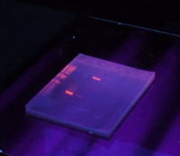
Effects of Ethidium Bromide: Ethidium bromide is a fluorescent dye that intercalates between bases of nucleic acids and allows very convenient detection of DNA fragments in gels, as shown by all the images on this page. As described above, it can be incorporated into agarose gels, or added to samples of DNA before loading to enable visualization of the fragments within the gel. As might be expected, binding of ethidium bromide to DNA alters its mass and rigidity, and therefore its mobility.

