\Experimental Steps
From 2007.igem.org
(Difference between revisions)
(→Our Preliminary Results) |
(→Step 2: Characterize Amplifier) |
||
| (One intermediate revision not shown) | |||
| Line 137: | Line 137: | ||
Clearly, the amplifier did not work the way we had thought it would. This highlights the importance of part characterization. If synthetic biology is going to advance, parts in the registry must be well characterized to be useful for engineering purposes. We took it upon ourselves to characterize this part to ensure that others would not run into the same problems we did. | Clearly, the amplifier did not work the way we had thought it would. This highlights the importance of part characterization. If synthetic biology is going to advance, parts in the registry must be well characterized to be useful for engineering purposes. We took it upon ourselves to characterize this part to ensure that others would not run into the same problems we did. | ||
| + | ====Experiments==== | ||
| + | [http://partsregistry.org/Part:BBa_J37015:Experience The following information can also be found in the Registry.] | ||
| - | |||
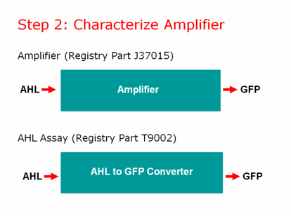
| + | Part J37015 was tested by the Brown iGEM team in August 2007. Some interesting and very repeatable results were obtained. It was expected that this part, J37015, would produce significantly more GFP than the part T9002, which differs only in that it has no LuxI gene and therefore no positive feedback loop. It was also expected that higher amounts of AHL would cause J37015 to produce more GFP than low amounts of AHL. Both of these hypotheses were proven false. | ||
| + | |||
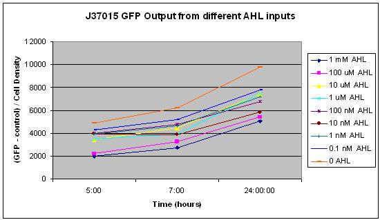
| + | In this experiment, 10 uL of a solution of AHL in water was mixed with 90 uL of LB and 3 uL of an overnight culture of cells. The concentrations 0, 0.1 nM, 1 nM, 10 nM, 100 nM, 1 uM, 10 uM, 100 uM, and 1 mM AHL were tested. These are the final concentrations of AHL in the solution with LB and cells, not the concentration of AHL in the water that was added to that solution. Measurements were made using a NanoDrop Spectrophotometer for Cell Density and a NanoDrop Flourospectrometer for GFP fluorescence at several time points over a 24 hour period. The average of all GFP measurements at the first measurement, time 0, was considered background fluorescence, and was subtracted from all subsequent GFP measurements. Additionally, all GFP measurements were divided by the cell density so that GFP per cell could be measured. This was to prevent noise from potential effects of AHL concentration on cell growth. | ||
| + | |||
| + | The first graph shows GFP output at 5, 7, and 24 hours after addition of AHL to the cells for each of the concentrations listed above. This clearly shows an inverse relationship between concentration of AHL and GFP output. This is quite surprising, because it was expected that more AHL would lead to more GFP expression. We do not know why this occurs, and are studying it further. Also, the fact that there is so much GFP expression when no AHL is added to the system indicates that this promoter acts at some constitutive level, and therefore has no use in a signal amplification system because it will always produce a signal regardless of its input. | ||
| + | |||
| + | [[Image:GFP_at_5,_7,_24_hours.JPG]] | ||
| + | |||
| + | |||
| + | |||
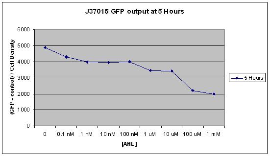
| + | The second graph shows the same result as the first, but looks only at the 5 hour time point. All time points after the first 2 hours showed indicated the inverse relationship between AHL concentration and GFP production described above, but it is most clear at 5 hours. | ||
| + | |||
| + | [[Image:5_hours.JPG]] | ||
| + | |||
| + | |||
| + | |||
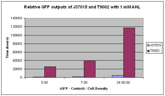
| + | The third graph shows a different result. The same concentration of AHL, 1 mM, was added to both J37015 and T9002. The only difference between these parts is that J37015 has the LuxI gene between its pLux promoter and its GFP gene. This theoretically creates a positive feedback loop, which should drive the cell to produce as much GFP as possible when it encounters any AHL. However, T9002 clearly had far more GFP output than J37015. This is even more intriguing when you consider the fact that T9002 has a weaker ribosome binding site for GFP than J37015 by 3 fold. One possible explanation for this is that the GFP gene is further from the promoter in J37015 than in T9002 due to the insertion of LuxI. However, it seems unlikely that this could cause such a strong difference. | ||
| + | |||
| + | [[Image:J37015_compared_to_T9002.JPG]] | ||
| + | |||
| + | |||
| + | |||
| + | From these results, it appears that J37015 does seem to have some sort of function, though not as a signal amplification device. Further investigation will hopefully reveal the cause of these unexpected data. | ||
==Step 3: Promoter and Lead Binding Protein== | ==Step 3: Promoter and Lead Binding Protein== | ||
Latest revision as of 02:29, 27 October 2007

