Tianjin/FLIP-FLOP/Model11
From 2007.igem.org
Sunlovedie (Talk | contribs) |
|||
| (4 intermediate revisions not shown) | |||
| Line 1: | Line 1: | ||
{| | {| | ||
| + | |- | ||
| + | |width="960px" style="padding: 10px; background-color: #FFFF99" | | ||
| + | <table width="99%" cellpadding="0" cellspacing="10" style="padding: 10px; background-color: #FFFF99"> | ||
| + | <tr> | ||
| + | <td><center>[[Image:tjumodel11a.jpg|350px]]<br><br> | ||
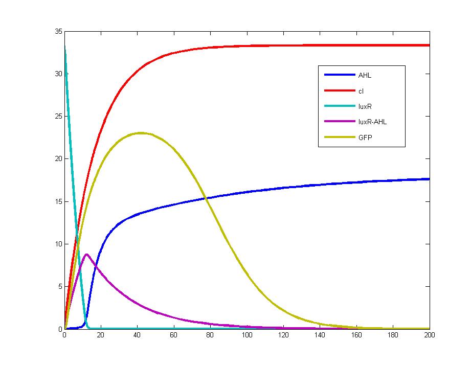
| + | Figure 1: This figure shows the concentration variation of chemical molecules when the input signal is at positive edge when t=0.<br></center></td> | ||
| + | <td><center>[[Image:tjumodel11b.jpg|350px]]<br><br> | ||
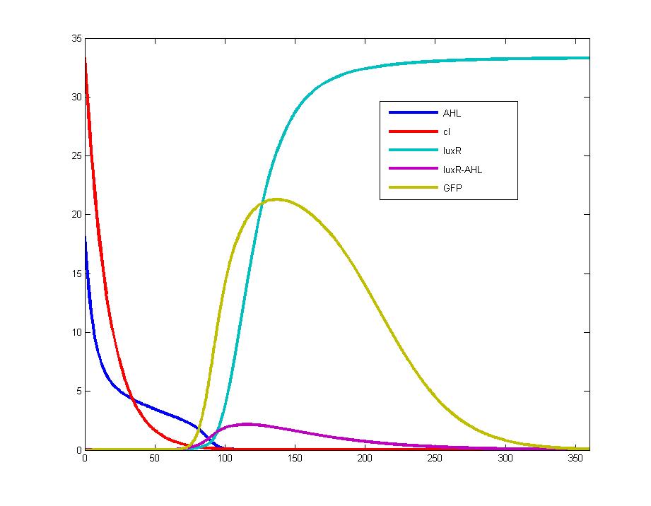
| + | Figure 2: This figure shows the concentration variation of chemical molecules when the input signal is at negative edge when t=0.<br></center></td> | ||
| + | </tr></table><br> | ||
These two pictures are drawn with the same parameters coming from related literature. Both figures point out that the output signal (GFP, yellow line) will form a pulse immediately after the input signal alters. | These two pictures are drawn with the same parameters coming from related literature. Both figures point out that the output signal (GFP, yellow line) will form a pulse immediately after the input signal alters. | ||
| - | <br> | + | <br><br> |
| - | + | ||
| - | + | ||
| - | + | ||
| - | + | ||
| - | + | ||
| - | Figure 3: This graph shows the variation of AHL, cI, LuxR and GFP responding to the addition of input | + | <table width="99%" cellpadding="0" cellspacing="10" style="padding: 10px; background-color: #FFFF99"> |
| - | signal(IPTG). It is easy to find that the | + | <tr> |
| - | with that of IPTG, whereas the contents of LuxR protein | + | <td>[[Image:TJUMODELFF203.jpg|400px]]<br></td> |
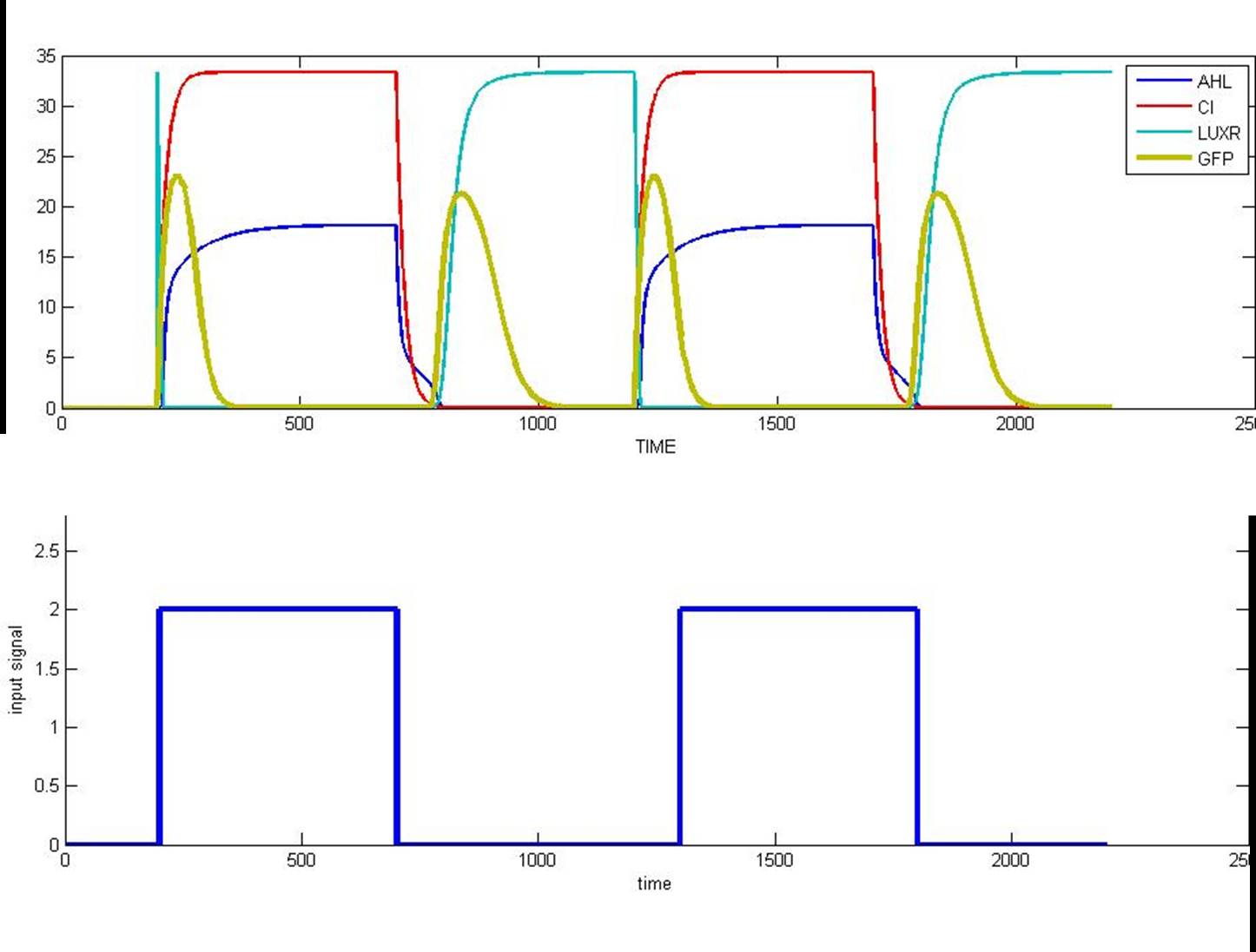
| - | Therefore, the output signal (GFP) only exists at the edge of IPTG curves where the input signal | + | <td>Figure 3: This graph shows the variation of AHL, cI, LuxR and GFP responding to the addition of input signal (IPTG). It is easy to find that the levels of AHL and cI share the same changing tendency with that of IPTG, whereas the contents of LuxR protein change inversely with that of IPTG. Therefore, the output signal (GFP) only exists at the edge of IPTG curves where the input signal switches from 1 to 0 or from 0 to 1. |
| - | switches from 1 to 0 or from 0 to 1. | + | </td> |
| - | + | </tr></table> | |
| - | + | ||
|} | |} | ||


