October: Gel Electrophoresis Report
From 2007.igem.org
SilvanaSaieh (Talk | contribs) m (October: Report on Electrophoresis moved to October: Gel Electrophoresis Report) |
SilvanaSaieh (Talk | contribs) |
||
| Line 10: | Line 10: | ||
| - | [[Image:Electro 1.jpg]] | + | [[Image:Electro 1.jpg|500px]] |
Revision as of 01:48, 24 October 2007
This is part of the experiments carried out in order to assemble different parts from the MIT BioBrick-Registry and those designed by the Colombian IGEM group. Thus, we could develop our chasses and devices.
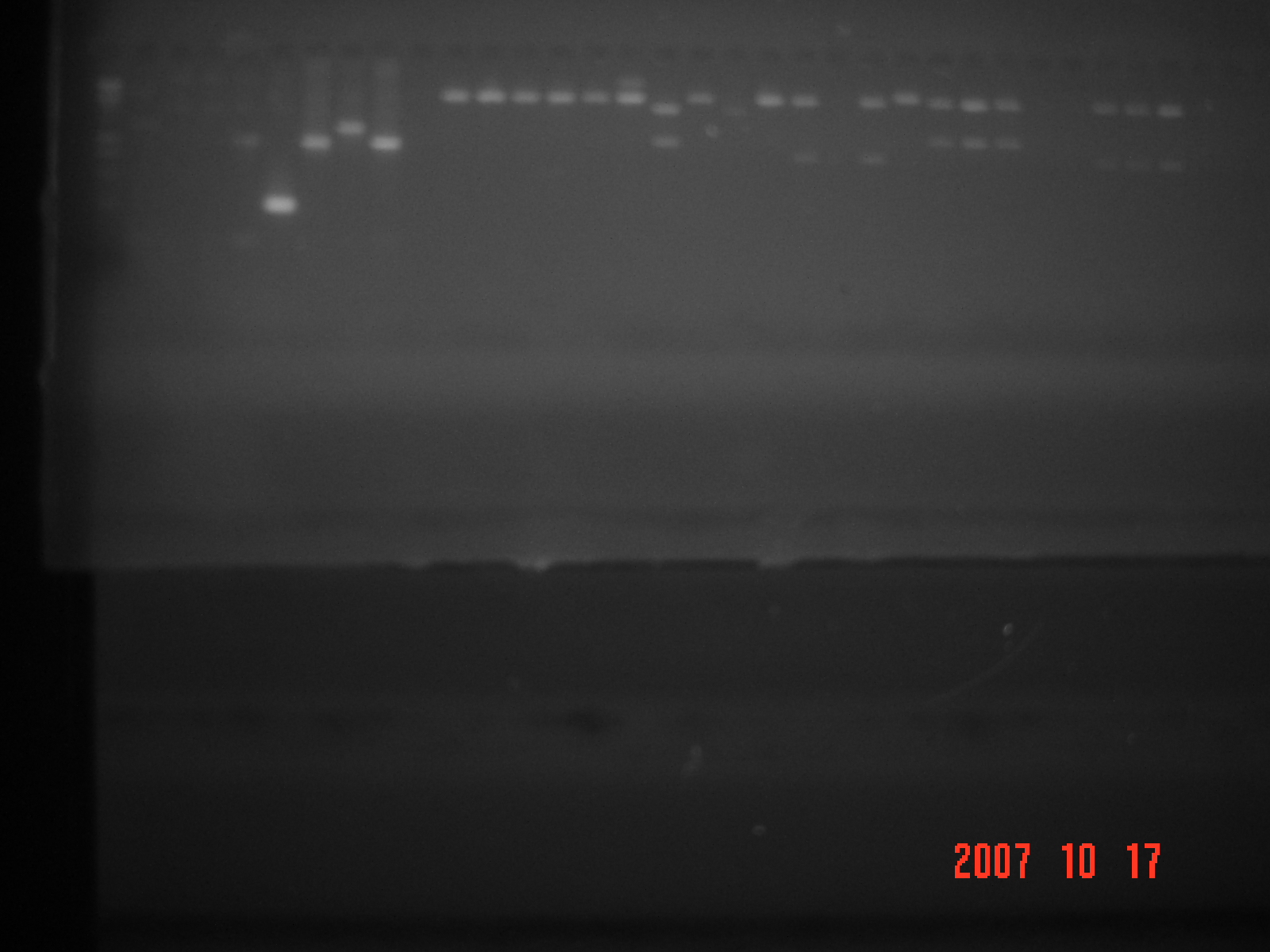
These are the results of the cutting of the parts. Electrophoresis ran on October 17, 2007.
On October 16 we performed PCR to amplify the extracted plasmids from our transformations. At the same time we did the necessary cuts using restriction enzymes XbaI and SpeI to separate the parts we were interested in from the plasmids, in addition we loaded them linearly so that they end up with the cohesive ends free where we could join the sequences of our interest.
Ahead we will present the first electrophoresis that we ran. In this picture you can see the plasmids extracted from the chemically transformed cells, the parts from the BioBrick and the sequences we will be incorporating.
Electrophoresis 1: Plasmids extracted from transformed cells
Lane
1. Control 1Kb
2. BioBrick part 9 O 2135 bp
3. BioBrick part 9 O 2135 bp
4. BioBrick part 4 P 3078 bp
5. BioBrick part 4 P 3078 bp
6. BioBrick part 11 A 2957 bp
7. BioBrick part 11 A 2957 bp
8. BioBrick part 13 D 3056 bp
9. BioBrick part 13 D 3056 bp
10. BioBrick part 8 E
11. BioBrick part 8 E
12. UV Promoter 2981 bp
13. UV Promoter 2981 bp
14. Exbb 3643 bp
15. Exbb 3643 bp
16. Fe Promoter 3951 bp
17. Fe Promoter 3951 bp
18. TomB 3641 bp
19. TomB 3641 bp
In this electrophoresis we found that the transformations were successful since you can see several bands with the expected size according to the plasmids used. Additionally, by duplicating them, we can confirm our results. Except for the bands of the parts 9 O and 8 E which aren’t where we expected them to be, all the transformations were successful. Because of this, we decided to not use the aforementioned parts in the upcoming experiments.
Following this test, we performed more extractions from these plasmids, and we carried out PCR on the chosen plasmids, doing the necessary cuts with the enzymes Xbal and SpeI.
Electrophoresis 2: Cuts from the plasmids and products of the PCR
Lane
1. Control 1 Kb
2. PCR 9 O
3. PCR 4 P
4. PCR 11 A
5. PCR 13 D
6. PCR Promoter UV
7. PCR Exbb
8. PCR Promoter Fe
9. PCR TomB
10. Space
11. 4 P cut by SpeI
12. 4 P cut by XbaI
13. 11 A cut by XbaI
14. 11 A cut by XbaI and SpeI
15. 11 A cut by XbaI
16. 13 D cut by XbaI
17. 13 D cut by XbaI and speI
18. Promoter UV cut by XbaI and SpeI
19. Promoter UV cut by XbaI
20. Promoter UV cut by XbaI and SpeI
21. Exbb cut by XbaI and SpeI
22. Space
23. Exbb cut by XbaI and SpeI
24. Exbb cut by XbaI
25. Promoter Fe cut by XbaI and SpeI
26. Promoter Fe cut by XbaI and SpeI
27. Promotor Fe cut by XbaI and SpeI
28. Space
29. Space
30. TomB cut by XbaI and SpeI
31. TomB cut by XbaI and SpeI
32. TomB cut by XbaI and SpeI
Now we will be comparing this last electrophoresis we the one we previously did starting from the last two lanes, starting from right to left.
In the lanes 30, 31, and 32 there are the digestions of the plasmid Psex which contains the encoded sequence for TomB; one of the proteins that makes up the transmembrane complex for the transport of iron-binding compounds called siderophores. The plasmid Psex with the incorporation of this sequence has a size of 3641 bp and the sequence by itself is 843 bp long. As you can see in the first picture, the migration of the complete circular plasmid matches the expected size of 3600 bp. In the second picture you can see the digestion of this plasmid with the two restriction enzymes; this produced two bands of different sizes. The uppermost band corresponds to the plasmid without the insertion of interest and a size of 2800 bp. The band which migrated the most, in other words, the band that can be found at the bottom of the gel corresponds to the sequence of TomB with cohesive ends cut by XbaI. (Take a look at the last three lanes.) The lanes of interest which contain the sequences for TomB were cut from the gel and were purified using the kit wizar Sv Gel and PCR Clean Up System. This procedure was carried out with all the sequences of our interest.
The lanes 27, 26, and 25 have the same procedure but this time with the intention of extracting the Fe Promoter. The complete plasmid has a size of 3951 bp and the insertion is of 1153 bp.
Lane 24 contains the plasmid Psex with the sequence that encodes for one of the other proteins that make up the transmembrane complex of siderophores called Exbb. In this lane we ran the lined-up plasmid with one cut made by XbaI. This procedure was carried out to utilize this plasmid as a vector for the protein previously mentioned with the UV Promoter once it has been incorporated. This way, we have the two sequences of our interest in one plasmid to be used in a bigger vector.
The lanes 23 and 21 correspond to this same plasmid with a double cut to extract the sequence of Exbb by itself. Therefore, you can observe a double band, unlike in lane 24.
In lanes 18, 19, and 20 are the plasmids Psex with the UV Promoters. In the lanes 28 and 20 we tried making a double cut to extract only the UV Promoter from the plasmid. Unfortunately the double band we were expecting in these lanes did not show up and we can only observe a single band.
Lanes 17 and 16 contain the sequence from the BioBrick 13 D. This sequence is very important for our device because it contains senders and receivers; in other words, it contains our upstream and downstream components. In the lane 17 you can observe two bands. The band that is in the lowest position contains the isolated part 13 D. This is a significant result because it will allow us to use this part numerous times. In the lane 16 you can observe this same plasmid with a single cut, lined up and ready to be joined with the necessary sequences. In this lane you can also notice a typical phenomenon of electrophoresis in which we can appreciate two bands where there should only be one. This comes about when one of the plasmids is more stretched and migrates less through the gel in comparison with the lined up plasmids which are more compact.
In the lane 15 you can see the BioBrick part 11 A lined up by a cut with restriction enzymes. This is an important part because it involves the upstream, downstream and where we will most probably set up our most complete device with all of our parts.
In the lanes 5, 6, 7, 8, and 9 are the products of the amplified PCR’s according to the previously mentioned convention.

