LarvaeSetupDish
From 2007.igem.org
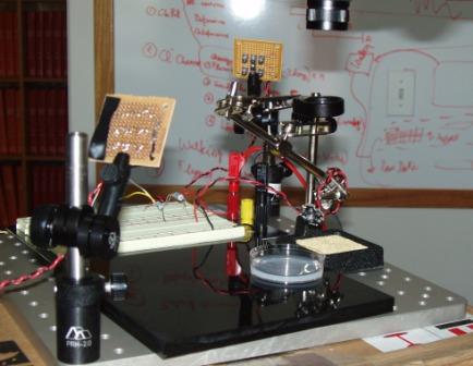
The apparatus includes a camera, a filter preventing visible light to pass through, a petri dish providing walking area for larvae, three infra red sources (2 at each side of the petri dish and 1 on top of the dish), a red light and a blue light. The distance from the light source (red/blue) to the center of the petri dish is 3.5cm. Larvae are taken one at a time to take the video. The diameter of the petri dish is 5 cm.
There are 3 types of videos taken for each strain. In type 1 (experiment A), 1s pulse of blue light followed by 6s recovery is done for 4 cycles. This is then followed by a long pulse of blue light. In type 2 experiment (experiment B), instead of 6 seconds between each pulse we wait until the larva recovers and starts moving normally again before activating the pulse. For each of these experiments, 10 trial runs are carried out.
From these videos, recovery time, extent of contraction, normal speed, and speed after recovery are measured and analysed.
In addition, in control experiments (type 3), 5 trial runs are carried out as in experment A but red light is used instead of blue light. For the last long pulse, the red light is kept on as long as the blue light in 5 experiments A. This is to confirm that the channel is not activated by red light. Hence, no response should be seen in larvae.
