Virginia Tech/phage
From 2007.igem.org
< Virginia Tech(Difference between revisions)
BlairLyons (Talk | contribs) m |
|||
| (6 intermediate revisions not shown) | |||
| Line 42: | Line 42: | ||
<img src="https://static.igem.org/mediawiki/2007/d/d5/Button_jc.JPG"/></a></html>[[Image:Dot1.JPG]] | <img src="https://static.igem.org/mediawiki/2007/d/d5/Button_jc.JPG"/></a></html>[[Image:Dot1.JPG]] | ||
| - | <!--sixth cell of second table: | + | <!--sixth cell of second table: Contributions and Contact--> |
| style="padding: 0px; width=50px; background-color: #FFFFFF;" | | | style="padding: 0px; width=50px; background-color: #FFFFFF;" | | ||
| - | <html><a href="https://2007.igem.org/Virginia_Tech/ | + | <html><a href="https://2007.igem.org/Virginia_Tech/Contributions"> |
| - | <img src="https://static.igem.org/mediawiki/2007/ | + | <img src="https://static.igem.org/mediawiki/2007/6/69/Button_contribute.JPG"/></a></html><html><a href="mailto:igem@vt.edu"></a></html> |
|}<html></center></html> | |}<html></center></html> | ||
| Line 72: | Line 72: | ||
| style="padding: 20px; background-color: #FFFFFF;" | | | style="padding: 20px; background-color: #FFFFFF;" | | ||
| - | <h3>Bacteriophage λ</h3> | + | [[Image:Fl_phage.PNG|center|]]<html><center></html><h3>Bacteriophage λ</h3><html></center></html> |
| - | λ phage is the viral component in our biological system that we used to test our model. λ phage has been characterized and studied for decades. | + | |
| - | + | λ phage is the viral component in our biological system that we used to test our model. We chose λ phage because it has been characterized and studied for decades. λ phage is also very stable and relatively easy to work with. We had a lot to learn since no one on the team had ever worked with λ phage before. | |
| - | + | ||
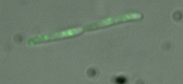
| + | At the beginning of the summer, we received fluorescent λ phage from the Laboratoire de Physique Statistique, Ecole Normale Supérieure, Paris, France. The phage was provided by Philippe Thomen and is characterized in <html><a href="https://static.igem.org/mediawiki/2007/3/32/Alvarez_2007.pdf">"Propagation of fluorescent viruses in growing plaques"</a></html> by Alvarez, L. J., P. Thomen, et al. Using this phage we were able to observe the build-up of viral particles in a lytic cell. | ||
| + | [[Image:Ecoli phage.PNG|thumb|center|400px|'''''E.coli'' infected with fluorescent phage.''' This cell is lytic: the green spots are new phage particles being constructed prior to lysis.]] | ||
|}<html></center></html> | |}<html></center></html> | ||
Latest revision as of 03:22, 27 October 2007
|
|
Bacteriophage λλ phage is the viral component in our biological system that we used to test our model. We chose λ phage because it has been characterized and studied for decades. λ phage is also very stable and relatively easy to work with. We had a lot to learn since no one on the team had ever worked with λ phage before. At the beginning of the summer, we received fluorescent λ phage from the Laboratoire de Physique Statistique, Ecole Normale Supérieure, Paris, France. The phage was provided by Philippe Thomen and is characterized in "Propagation of fluorescent viruses in growing plaques" by Alvarez, L. J., P. Thomen, et al. Using this phage we were able to observe the build-up of viral particles in a lytic cell.
|


