Imperial/Wet Lab/Results/CBD1.1
From 2007.igem.org
(→Conclusion) |
m |
||
| (9 intermediate revisions not shown) | |||
| Line 1: | Line 1: | ||
{{Template: IC07navmenu}} | {{Template: IC07navmenu}} | ||
| + | <br clear="all"> | ||
__NOTOC__ | __NOTOC__ | ||
| Line 22: | Line 23: | ||
| width="200px"| [[Image:IC2007 Experiments Phase1 Protocol1-1.jpg|thumb|300px|Fig.2: pTet ''in vivo'' over 6 hours]] | | width="200px"| [[Image:IC2007 Experiments Phase1 Protocol1-1.jpg|thumb|300px|Fig.2: pTet ''in vivo'' over 6 hours]] | ||
| width="50px"| | | width="50px"| | ||
| - | |width="300px"| pTet-GFP and pT7-GFP were both compared to a negative and positive control, where pTet-GFP showed significant fluorescence, while pT7 did not show any dramatic increase (Fig.1). When measured over intervals, pTet showed a steady increase in fluorescence (Fig.2). Fig.2 also suggests that there may be some variation across the samples that have been measured. | + | |width="300px"| pTet-GFP and pT7-GFP were both compared to a negative and a positive control, where pTet-GFP showed significant fluorescence, while pT7 did not show any dramatic increase in fluorescence (Fig.1). When measured over intervals, pTet showed a steady increase in fluorescence (Fig.2). Fig.2 also suggests that there may be some variation across the samples that have been measured. |
|} | |} | ||
<br clear=all> | <br clear=all> | ||
| Line 31: | Line 32: | ||
| width="200px"| [[Image:IC2007 Experiments Phase1 Protocol1-1Res1.PNG|thumb|310px|Fig.4 - pT7-GFP ''in vivo'' over 4 hours]] | | width="200px"| [[Image:IC2007 Experiments Phase1 Protocol1-1Res1.PNG|thumb|310px|Fig.4 - pT7-GFP ''in vivo'' over 4 hours]] | ||
| width="50px"| | | width="50px"| | ||
| - | |width="300px"| Fig.3 shows that the pT7-GFP construct had significant fluorescence compared to the negative control, which seems to suggest that GFP has been expressed. However, when fluorescence | + | |width="300px"| Fig.3 shows that the pT7-GFP construct had significant fluorescence compared to the negative control, which seems to suggest that GFP has been expressed. However, when fluorescence was plotted against time, there was little or no increase in fluorescence (Fig.4), suggesting little or no expression of GFP over the time course. |
|} | |} | ||
<br clear="all"> | <br clear="all"> | ||
| Line 38: | Line 39: | ||
*Positive control - diluted GFP solution of equal volume | *Positive control - diluted GFP solution of equal volume | ||
*Negative control - LB media of equal volume | *Negative control - LB media of equal volume | ||
| + | |||
| + | [[Media:IC 2007 PT7 in vitro 37oC.xls|Excel file of Raw data]] | ||
| Line 51: | Line 54: | ||
===[http://partsregistry.org/Part:BBa_E7104 '''pT7-GFP''']=== | ===[http://partsregistry.org/Part:BBa_E7104 '''pT7-GFP''']=== | ||
| - | As our initial tests of the pT7-GFP construct did not yield expected results, it was postualated that the absence of fluorescence increase could be due to several reasons. Firstly, the cells might not be properly induced, to which IPTG of a suitable concentration(~1mM) needs to be added, and the culture grown overnight. It could also be that the construct was not working at all, or that the culture was in stationary phase when it was tested, leading to little or | + | As our initial tests of the pT7-GFP construct did not yield expected results, it was postualated that the absence of fluorescence increase could be due to several reasons. Firstly, the cells might not be properly induced, to which IPTG of a suitable concentration (~1mM) needs to be added, and the culture grown overnight. It could also be that the construct was not working at all, or that the culture was in stationary phase when it was tested, leading to little or no expression of GFP. |
| - | + | ||
| - | + | ||
| + | In the re-test, the culture had been properly induced with IPTG at growth phase. While absolute fluorescence levels indicate the presence of GFP in the culture, it was not as significant as that of pTet-GFP. In addition, there was little or no increase of fluorescence over the time period at which it was measured. This could be due to the fact that the pT7-GFP construct either did not express as quickly or strongly as pTet-GFP ''in vivo'', or that the construct was simply not working. Further investigation into the construct also revealed a weaker ribosome binding site compared to the pTet-GFP construct which could account for the weak fluorescent signal. | ||
==Conclusion== | ==Conclusion== | ||
We investigated the possibility of using different constructs, and strength of different promoters for our projects. To conclude, | We investigated the possibility of using different constructs, and strength of different promoters for our projects. To conclude, | ||
* pTet-GFP construct gave a strong fluorescent signal, indicating good expression of GFP ''in vivo''. | * pTet-GFP construct gave a strong fluorescent signal, indicating good expression of GFP ''in vivo''. | ||
| - | * pT7-GFP construct gave little or no fluorescent signal, to which several reasons had been suggested, and further experiments are required to | + | * pT7-GFP construct gave little or no fluorescent signal, to which several reasons had been suggested, and further experiments are required to confirm them. |
| - | + | ||
| - | + | ||
Latest revision as of 02:21, 27 October 2007

In vivo Testing of pTet-GFP and pT7-GFP Constructs
Aims
To determine if the following constructs work in vivo:
- [http://partsregistry.org/Part:BBa_I13522 pTet-GFP]
- [http://partsregistry.org/Part:BBa_E7104 pT7-GFP]
The testing was comprised of several tests:
- pTet and pT7 in vivo - Tested 16-08-2007
- Retesting pT7 in vivo - Tested 17-08-2007
Materials and Methods
Link to the Protocol
Results
Test: 16-08-2007
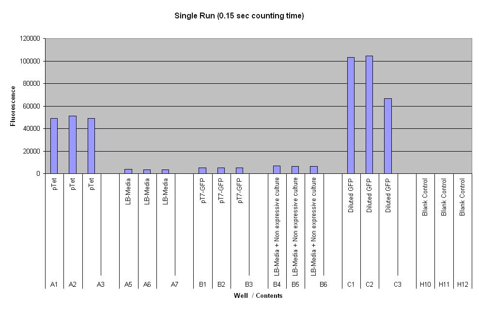
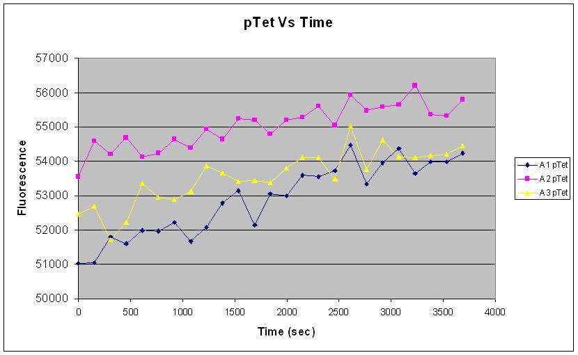
| pTet-GFP and pT7-GFP were both compared to a negative and a positive control, where pTet-GFP showed significant fluorescence, while pT7 did not show any dramatic increase in fluorescence (Fig.1). When measured over intervals, pTet showed a steady increase in fluorescence (Fig.2). Fig.2 also suggests that there may be some variation across the samples that have been measured. |
Test: 17-08-2007
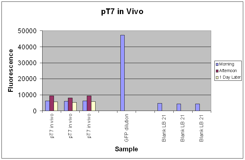
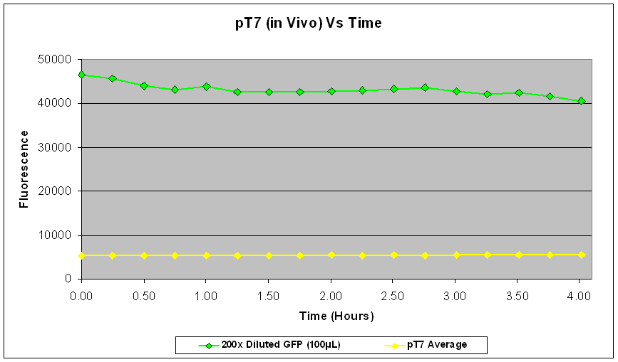
| Fig.3 shows that the pT7-GFP construct had significant fluorescence compared to the negative control, which seems to suggest that GFP has been expressed. However, when fluorescence was plotted against time, there was little or no increase in fluorescence (Fig.4), suggesting little or no expression of GFP over the time course. |
Controls:
- Positive control - diluted GFP solution of equal volume
- Negative control - LB media of equal volume
Discussion
[http://partsregistry.org/Part:BBa_I13522 pTet-GFP]
Fig.1 and Fig.2 both strongly indicate that there was a good amount of expression of GFP (~54000) with the pTet-GFP construct, leading to an increase in fluoresence over time. This shows that the construct is functioning well in vivo.
Fig.2 however suggests that there may be significant variation in the measurement of our samples. This could be due to experimental methodology, or the intrinsic nature of variability of expression in the in vivo chassis.
[http://partsregistry.org/Part:BBa_E7104 pT7-GFP]
As our initial tests of the pT7-GFP construct did not yield expected results, it was postualated that the absence of fluorescence increase could be due to several reasons. Firstly, the cells might not be properly induced, to which IPTG of a suitable concentration (~1mM) needs to be added, and the culture grown overnight. It could also be that the construct was not working at all, or that the culture was in stationary phase when it was tested, leading to little or no expression of GFP.
In the re-test, the culture had been properly induced with IPTG at growth phase. While absolute fluorescence levels indicate the presence of GFP in the culture, it was not as significant as that of pTet-GFP. In addition, there was little or no increase of fluorescence over the time period at which it was measured. This could be due to the fact that the pT7-GFP construct either did not express as quickly or strongly as pTet-GFP in vivo, or that the construct was simply not working. Further investigation into the construct also revealed a weaker ribosome binding site compared to the pTet-GFP construct which could account for the weak fluorescent signal.
Conclusion
We investigated the possibility of using different constructs, and strength of different promoters for our projects. To conclude,
- pTet-GFP construct gave a strong fluorescent signal, indicating good expression of GFP in vivo.
- pT7-GFP construct gave little or no fluorescent signal, to which several reasons had been suggested, and further experiments are required to confirm them.


