Imperial/Wet Lab/Lab Notebook/2007-08-21
From 2007.igem.org
< Imperial | Wet Lab | Lab Notebook(Difference between revisions)
m |
|||
| (3 intermediate revisions not shown) | |||
| Line 1: | Line 1: | ||
| + | __NOTOC__ | ||
{{Template:IC07navmenu}} | {{Template:IC07navmenu}} | ||
<html><div id="maincol"></html> | <html><div id="maincol"></html> | ||
| Line 4: | Line 5: | ||
= 21 August 2007 = | = 21 August 2007 = | ||
| + | ==Cloning of Biobricks== | ||
| + | #Placed 5 ml of LB in a 15 ml tube | ||
| + | #Added appropriate antibiotics into the tube | ||
| + | #Picked a colony from the fresh overnight plate | ||
| + | #Innoculated the colony in the LB media | ||
| + | #Incubated for 6 hours during the day | ||
| + | #*2x5 Biobricks cloned | ||
| + | |||
| + | ==Cloning of Biobricks (for Midiprep)== | ||
| + | #Placed 50 ml of LB in a 250 ml conical flask | ||
| + | #Innoculated 1 ml of culture into LB media in flask | ||
| + | #Incubated overnight at 37 °C | ||
| + | |||
| + | *<font size=-2>BBa_J23039 [tet-luxI] | ||
| + | *BBa_I13504 [GFP] | ||
| + | *BBa_R0062 [pLux (no luxR)] | ||
| + | *BBa_J37032 [pLux-GFP] | ||
| + | *BBa_S03119 [tet-LuxR]</font> | ||
| + | |||
| + | ==Tasks Completed== | ||
| + | #Measured the fluorescence of pT7 in vivo. | ||
| + | #Measured the fluorescence of pTet in vitro over a time span of 6 hours, at hourly intervals. | ||
| + | #Tested the effects of fluorescence window on the variation of fluorescence readings and degradation of GFP. | ||
| + | #Did the initial testing of calibration of GFP- dilution of the same [GFP] to different total volumes. | ||
| + | |||
| + | ==Aims== | ||
| + | #Protocol 1- In vivo testing for pT7 test construct | ||
| + | #Protocol 2- In vitro testing in commercial cell extract for pTet, pT7, pLux test constructs | ||
| + | #Protocol 3- Fluorescence dependence protocol, this is an initial test before the calibration curve and will affect how this is designed. | ||
| + | #Protocol 4- Fluorometer optimization, this is a protocol to try minimize the variation that we saw in the initial in vivo tests | ||
| + | |||
| + | ===Protocol 1=== | ||
| + | ===Protocol 2=== | ||
| + | '''Aims:''' | ||
| + | *To test pTet ''in vitro' | ||
| + | ====Equipment==== | ||
| + | *Fluorometer + Connected PC '''Turn on before beginning''' | ||
| + | *37<sup>o</sup>C | ||
| + | *96 well plate x1 + Plate lid + Autoclave tape | ||
| + | *1.5ml eppendorf tube x7 | ||
| + | *eppendorf rack | ||
| + | *Gilson p20,p200,p1000 | ||
| + | *Stop watch | ||
| + | |||
| + | ====Reagents==== | ||
| + | *Commercial S30 E.coli extract. <font color=green> Level 5 Biochem freezer</font> | ||
| + | **175µl Amino Acid Mixture Minus Methionine, 1mM | ||
| + | **175µl Amino Acid Mixture Minus Leucine, 1mM | ||
| + | ** 1 x 45µl tubes of S30 Extract, Circular | ||
| + | ** 1 x 60µl S30 Premix Without Amino Acids | ||
| + | *GFP solution (For this initial experiment does not need to be purified GFP, we just want to know we have the right filter and that our settings are adjusted to measuring GFP) <font color=green> In the teaching labs fridge </font> | ||
| + | *pTET midi DNA id 02<font color=green> In the teaching labs fridge</font> | ||
| + | |||
| + | ====Protocols==== | ||
| + | #First collect all equipment and reagents and ensure the fluorometer and that the PC connected has a data collection protocol installed. All the cell extracts are kept level 5 in the biochemistry labs in the bottom draw of the -80<sup>o</sup>C freezer. The S30 extract and premix have been divided into 10 tubes, 1 tube being enough for 3 reactions. The amino acid mixtures have not been divided and should be removed from the freezer and the volumes measured out in the biochemistry level 5 labs. | ||
| + | #''Commercial E.coli Cell Extract'': First prepare a complete amino acid mixture for both extract solutions: Add 7.5μl volume of two amino acid minus mixtures into an labeled eppendorf to give a volume of 15μl. Each amino acid minus mixture is missing one type of amino acid, and so by combining two solutions we are complementing each solution for the missing amino acid. Place eppendorf in a rack on bench. | ||
| + | #''Commercial E.coli Cell Extact'':Take an eppendorf tube and add 5µl of the E.coli complete amino acid mixture. Then add 20µl of S30 Premix Without Amino Acid. Then add 15µl of S30 Extract Circular. The volume should be 40ul in the eppendorf tube. | ||
| + | #Carry out step above twice more so that we end up with three eppendorf tubes of prepared commercial E.coli extract, however for one of the tubes mark as '''control''' and add 20ul of nuclease free water to the eppendorf. | ||
| + | #Vortex the tubes to mix thoroughly and place the 5x eppendorf tubes in the incubator at 37<sup>o</sup>C | ||
| + | |||
| + | '''Loading Plate''' | ||
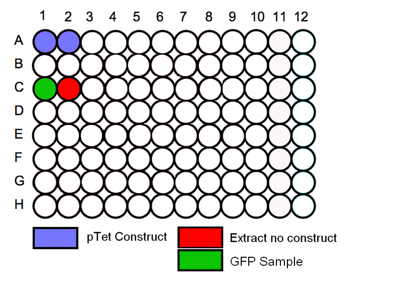
| + | #Follow the schematic for the plate and begin by loading the in vitro expression system into the correct wells. Before loading in the samples vortex the tubes for a few seconds to mix the solution. | ||
| + | #Place the lid on the 96well plate and tape up the edges of the lid. This should be put into the incubator at 37<sup>o</sup>C for 10 minutes to allow temperature to equilibrate | ||
| + | #Remove from 37<sup>o</sup>C incubator and spin-down in centrifuge in plate centrifuge at 2000rpm for a few seconds. Spin down is the process of bringing down any solution on lid or side of well into the base of the well. Alternatively can tap the top of the lid to bring down any solution to bottom of the well. | ||
| + | #'''CAREFULLY''' remove lid off the 96well plate and place in the fluorometer. Create a file name protocol 2-1 under: D:\IGEM\'''INSERT DATE'''\CBD\ protocol 2-1. Export the data here. If repeated measurements change the second number to suit repeat number, e.g. 2nd repeat protocol 2-2, 5th repeat protocol 2-5. Once the data collection is set up then initiate the measurements. | ||
| + | #This measurement will give a back ground fluorescence measurement and can be used as our time zero data. | ||
| + | #Then to begin the reaction add 20ul volume of purified DNA sample to give 2µg to the wells indicated on the schematic. Be careful not to add to wells that DO NOT NEED DNA or to mix up which DNA we are adding. | ||
| + | #Place lid back on and tape again and place back in the incubator at 37<sup>o</sup>C <br><br> | ||
| + | #After 30 minutes of incubation measure the fluorescence by repeating procedure 3-4 above. This initial measurement of 30 minutes is to find out how fast GFP is being produced. After this initial measurement, the intervals should be reassessed and adjusted accordingly | ||
| + | #Before each measurement be careful to remember to either spin down or tap down the solution and to remove the lid before placing in the fluorometer | ||
| + | |||
| + | '''Schematic''' | ||
| + | {| border="1" cellpadding="1" | ||
| + | | | ||
| + | {| border="1" cellpadding="2" | ||
| + | !<u>Well </u> || <u>Test Construct</u> !! <u>In vitro chassis</u> !! <u>Vol of in <br> vitro chassis (ul)</u> | ||
| + | |- | ||
| + | |<font color=blue> A1 | ||
| + | |<font color=blue> pTet (20ul) | ||
| + | |<font color=blue>Commercial E.coli extract | ||
| + | |<font color=blue>40 | ||
| + | |- | ||
| + | |<font color=blue>A2 | ||
| + | |<font color=blue>pTet (20ul) | ||
| + | |<font color=blue>Commercial E.coli extract | ||
| + | |<font color=blue>40 | ||
| + | |- | ||
| + | |<font color=green>C1 | ||
| + | |<font color=green>None | ||
| + | |<font color=green>GFP sample | ||
| + | |<font color=green>None | ||
| + | |- | ||
| + | |<font color=red>C2 | ||
| + | |<font color=red>None | ||
| + | |<font color=red>Control Commercial E.coli extract | ||
| + | |<font color=red>60 | ||
| + | |} | ||
| + | | | ||
| + | [[Image:IC07 21 08 2007 plate1.PNG|400px|96 Plate Schematic]] | ||
| + | |} | ||
| + | |||
| + | ==Pilot Preparation of Vesicles== | ||
| + | |||
| + | '''Preparations''' | ||
| + | |||
| + | The desiccation and suspension stages of the vesicle preparation protocol were followed. | ||
| + | * One 100ml beaker was prepared with 125μl of DOPC in 50ml of mineral oil. | ||
| + | * The suspension was sonicated for 30mins and left overnight in a 27°C incubator. | ||
<html></div></html> | <html></div></html> | ||
{{Template:IC07labnotebook}} | {{Template:IC07labnotebook}} | ||
Latest revision as of 23:59, 24 October 2007

21 August 2007
Cloning of Biobricks
- Placed 5 ml of LB in a 15 ml tube
- Added appropriate antibiotics into the tube
- Picked a colony from the fresh overnight plate
- Innoculated the colony in the LB media
- Incubated for 6 hours during the day
- 2x5 Biobricks cloned
Cloning of Biobricks (for Midiprep)
- Placed 50 ml of LB in a 250 ml conical flask
- Innoculated 1 ml of culture into LB media in flask
- Incubated overnight at 37 °C
- BBa_J23039 [tet-luxI]
- BBa_I13504 [GFP]
- BBa_R0062 [pLux (no luxR)]
- BBa_J37032 [pLux-GFP]
- BBa_S03119 [tet-LuxR]
Tasks Completed
- Measured the fluorescence of pT7 in vivo.
- Measured the fluorescence of pTet in vitro over a time span of 6 hours, at hourly intervals.
- Tested the effects of fluorescence window on the variation of fluorescence readings and degradation of GFP.
- Did the initial testing of calibration of GFP- dilution of the same [GFP] to different total volumes.
Aims
- Protocol 1- In vivo testing for pT7 test construct
- Protocol 2- In vitro testing in commercial cell extract for pTet, pT7, pLux test constructs
- Protocol 3- Fluorescence dependence protocol, this is an initial test before the calibration curve and will affect how this is designed.
- Protocol 4- Fluorometer optimization, this is a protocol to try minimize the variation that we saw in the initial in vivo tests
Protocol 1
Protocol 2
Aims:
- To test pTet in vitro'
Equipment
- Fluorometer + Connected PC Turn on before beginning
- 37oC
- 96 well plate x1 + Plate lid + Autoclave tape
- 1.5ml eppendorf tube x7
- eppendorf rack
- Gilson p20,p200,p1000
- Stop watch
Reagents
- Commercial S30 E.coli extract. Level 5 Biochem freezer
- 175µl Amino Acid Mixture Minus Methionine, 1mM
- 175µl Amino Acid Mixture Minus Leucine, 1mM
- 1 x 45µl tubes of S30 Extract, Circular
- 1 x 60µl S30 Premix Without Amino Acids
- GFP solution (For this initial experiment does not need to be purified GFP, we just want to know we have the right filter and that our settings are adjusted to measuring GFP) In the teaching labs fridge
- pTET midi DNA id 02 In the teaching labs fridge
Protocols
- First collect all equipment and reagents and ensure the fluorometer and that the PC connected has a data collection protocol installed. All the cell extracts are kept level 5 in the biochemistry labs in the bottom draw of the -80oC freezer. The S30 extract and premix have been divided into 10 tubes, 1 tube being enough for 3 reactions. The amino acid mixtures have not been divided and should be removed from the freezer and the volumes measured out in the biochemistry level 5 labs.
- Commercial E.coli Cell Extract: First prepare a complete amino acid mixture for both extract solutions: Add 7.5μl volume of two amino acid minus mixtures into an labeled eppendorf to give a volume of 15μl. Each amino acid minus mixture is missing one type of amino acid, and so by combining two solutions we are complementing each solution for the missing amino acid. Place eppendorf in a rack on bench.
- Commercial E.coli Cell Extact:Take an eppendorf tube and add 5µl of the E.coli complete amino acid mixture. Then add 20µl of S30 Premix Without Amino Acid. Then add 15µl of S30 Extract Circular. The volume should be 40ul in the eppendorf tube.
- Carry out step above twice more so that we end up with three eppendorf tubes of prepared commercial E.coli extract, however for one of the tubes mark as control and add 20ul of nuclease free water to the eppendorf.
- Vortex the tubes to mix thoroughly and place the 5x eppendorf tubes in the incubator at 37oC
Loading Plate
- Follow the schematic for the plate and begin by loading the in vitro expression system into the correct wells. Before loading in the samples vortex the tubes for a few seconds to mix the solution.
- Place the lid on the 96well plate and tape up the edges of the lid. This should be put into the incubator at 37oC for 10 minutes to allow temperature to equilibrate
- Remove from 37oC incubator and spin-down in centrifuge in plate centrifuge at 2000rpm for a few seconds. Spin down is the process of bringing down any solution on lid or side of well into the base of the well. Alternatively can tap the top of the lid to bring down any solution to bottom of the well.
- CAREFULLY remove lid off the 96well plate and place in the fluorometer. Create a file name protocol 2-1 under: D:\IGEM\INSERT DATE\CBD\ protocol 2-1. Export the data here. If repeated measurements change the second number to suit repeat number, e.g. 2nd repeat protocol 2-2, 5th repeat protocol 2-5. Once the data collection is set up then initiate the measurements.
- This measurement will give a back ground fluorescence measurement and can be used as our time zero data.
- Then to begin the reaction add 20ul volume of purified DNA sample to give 2µg to the wells indicated on the schematic. Be careful not to add to wells that DO NOT NEED DNA or to mix up which DNA we are adding.
- Place lid back on and tape again and place back in the incubator at 37oC
- After 30 minutes of incubation measure the fluorescence by repeating procedure 3-4 above. This initial measurement of 30 minutes is to find out how fast GFP is being produced. After this initial measurement, the intervals should be reassessed and adjusted accordingly
- Before each measurement be careful to remember to either spin down or tap down the solution and to remove the lid before placing in the fluorometer
Schematic
|
Pilot Preparation of Vesicles
Preparations
The desiccation and suspension stages of the vesicle preparation protocol were followed.
- One 100ml beaker was prepared with 125μl of DOPC in 50ml of mineral oil.
- The suspension was sonicated for 30mins and left overnight in a 27°C incubator.
